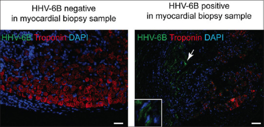
Figure 3.

Representative images showing Immunofluorescence analysis of HHV-6B specific staining in cardiac allograft tissues using HHV-6B OHV-3 antibody. Troponin (red) staining was used as a control for cardiac tissue specific staining. HHV-6B positive areas (green) are indicated with white arrowhead. Expanded image of HHV-6B positive staining is shown within a white box. The scale bar represents 10 μm
