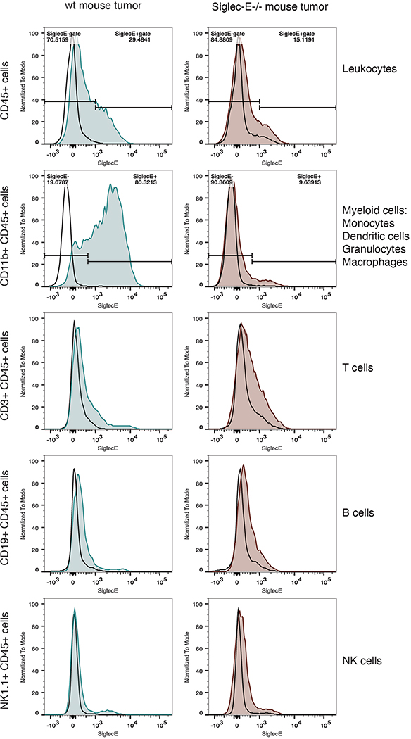
Extended Data Fig. 10 |. B16D5 tumors in C57Bl/6 mice have significant infiltration of Siglec-E+ immune cells, which can largely be attributed to Siglec-E expressing CD11b+ leukocytes.
Representative histograms of tumor infiltrating leukocytes gated as shown in Supplementary Fig. 22, with Siglec-E expression on the biexponential x-axis and cell count normalized to mode (n = 10 for wt mice, n = 12 for Siglec-E deficient mice). A fluorescence minus one control (FMO, negative control that lacks the Siglec-E antibody) for each sample is shown as a black outline. Total leukocytes (defined as CD45+ cells) show a detectable increased Siglec-E expression in wild-type mice (blue) compared to Siglec-E−/− mouse controls (red). Investigating further, T cells, B cells, and NK cells (represented by CD3+, CD19+, and NK1.1+ leukocyte populations did not show significant Siglec-E expression compared to Sig-E−/− control cells, however, myeloid (CD11b+) cells, which include macrophages, granulocytes, certain dendritic cells, and monocytes, had significant anti Siglec-E signal and represent the vast majority of Siglec-E+ leukocytes in these tumors.

