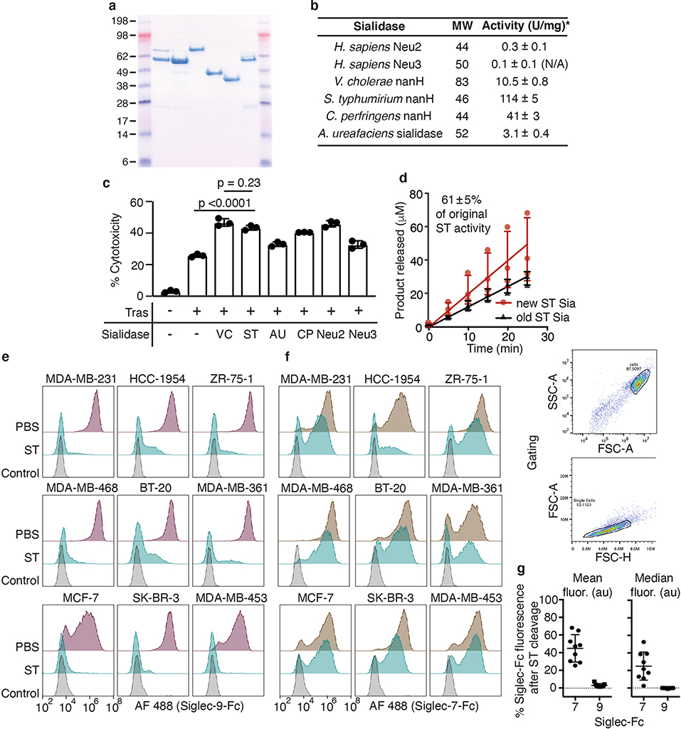
Extended Data Fig. 2 |. ST sialidase is a small, stable sialidase that enhances NK cell-mediated ADCC towards breast cancer cells and cleaves Siglec-7 and -9 ligands at high concentrations.
a, PAGE-SDS reducing gel of six recombinantly expressed and purified sialidases: Neu2, Neu3, Vibrio cholerae sialidase, Salmonella typhimurium NanH, Clostridium perfringens NanH, Arthrobacter ureafaciens NanH b, Molecular weights and specific activities (in μmol substrate converted per min per mg enzyme) of sialidases determined by in vitro activity assays with the fluorogenic substrate 4-MUNANA (n = 3 experimental replicates; mean ± SD). c, IL2-activated NK cell-mediated ADCC assay against target BT-20 breast cancer cells treated with 10 nM trastuzumab (Tras) and 2 μM sialidases at E/T = 4, percent cytotoxicity was detected by LDH release after 4 h. Figure shows mean ± SD from n = 3 experimental replicates against the same biological NK cell donor. Statistical analysis by one way ANOVA with Tukey’s multiple comparisons adjusted p-values. d, In vitro activity assay of freshly expressed sialidase compared with sialidase stored in PBS at 4 °C for 2 years reveals that significant ST sialidase activity is preserved (n = 3 experimental replicates fit to a linear regression ± SD). e-g, Siglec ligand depletion by ST sialidase cleavage is effective across many breast cancer cell lines. ST sialidase (2 μM) was incubated with nine different breast cancer cell lines for 1 h and the removal of Siglec ligands compared to PBS-treated cells was assessed by staining with Siglec-9-Fc (e) or Siglec-7-Fc (f) and anti-human-488 secondary antibody (control: secondary only). Gating is shown on the right, first selecting the cell population (FSC-A/SSC), then gating on single cells (FSC-A/FSC-H). Representative images from n = 2 experimental replicates of the n = 9 biological cell line replicates each with >2,000 cells are shown. The mean and median (± SD) percent decrease in Siglec ligand-binding fluorescent signal upon ST treatment of the n = 9 cell lines are quantified in g.

