
Figure 1.
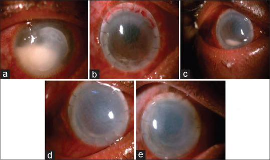
(a) Day of Presentation, 5 × 6 mm infiltrate with underlying endothelial plaque and 4.0 mm hypopyon. (b) PostOP Day 1 with intact graft host junction with minimal hypopyon. (c) 3rd post op week showing graft edema with resolving hypopyon. (d) 4 th post operative week showing resolving hypopyon. (e) 2 nd month post operative showing failed graft with resolved hypopyon