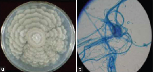
Figure 2.

(a) Growth of M. wolfii on SDA. Growth of M. wolfii on SDA showing grayish white, downy, with a broad zonated rosette- like colony. (b) Morphology of M. wolfii in Lactophenol cotton blue staining. LPCB under × 40 magnification showing the sterile mycelia (Non Sporulating Mould (NSM))
