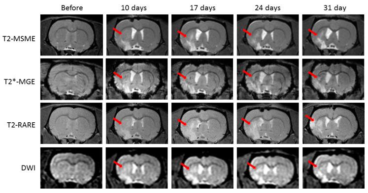
Figure 1.
Example of the ischemic lesion evolution detected in the rat brain In vivo before middle-cerebral-artery occlusion (MCAO) and on days 10, 17, 24, and 31 after surgery and days 7, 14, 21, and 28 after phosphate-buffered saline (PBS) injection on T2-weighted multislice multiecho (T2-MSME), T2*-weighted multiple gradient echo (T2*-MGE), T2-weighted turbo rapid acquisition with relaxation enhancement (T2 TURBO RARE), and diffusion-weighted images (DWI). Red arrows point to the ischemic lesion area.

