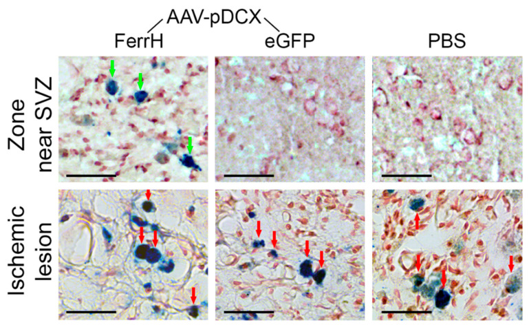
Figure 6.
Perl’s Prussian blue histological staining for detection of iron accumulation in the zone near lateral ventricle and SVZ (top row) and in the ischemic lesion zone (bottom row). Green arrows point to the cells with iron accumulation in the zone near the SVZ; red arrows point to the cells with iron accumulation in the ischemic lesion. Scale bar corresponds to 50 µm. Abbreviations: AAV, adeno-associated viral backbone; pDCX, doublecortin promoter; FerrH, ferritin heavy chain; eGFP, enhanced green fluorescent protein; PBS, phosphate-buffered saline; SVZ, subventricular zone.

