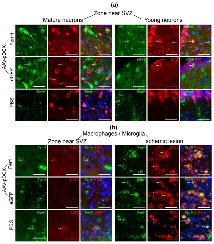
Figure 7.
Examples of immunofluorescent staining for FerrH, NeuN (mature neurons), DCX (young neurons), and activated microglia\macrophages (CD68). (a) Double-immunofluorescence images stained for mature (FerrH\NeuN for the Ferr-MCAO and PBS-MCAO group, eGFP\NeuN for the eGFP-MCAO group), young (FerrH\DCX for the FerrH-MCAO and PBS-MCAO group, and eGFP\DCX for the eGFP-MCAO group) neurons in the zone near the lateral ventricle and SVZ. (b) Double-immunofluorescence images stained for microglia\macrophages (FerrH\CD68) in the near lateral ventricle and SVZ (left three rows) and inside the ischemic lesion (right three rows). For eGFP-MCAO group, FerrH is visualized by far-red fluorochrome and is shown as green pseudo color. Scale bars corresponds to 50 µm. Abbreviations: MCAO, middle-cerebral-artery occlusion; AAV, adeno-associated viral backbone; pDCX, doublecortin promoter; FerrH, ferritin heavy chain; eGFP, enhanced green fluorescent protein; PBS, phosphate-buffered saline; SVZ, subventricular zone.

