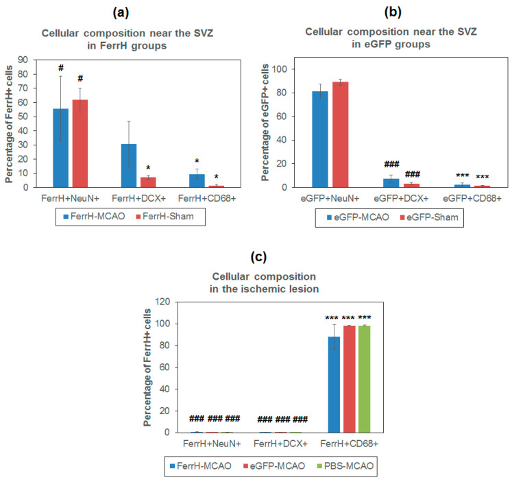
Figure 8.
Cellular composition of two zones of MRI signal hypointensity in T2*-weighted images, near the SVZ and inside of the ischemic lesion area. (a) Percentage of mature (NeuN+) and immature (DCX+) neurons and activated microglia\macrophages (CD68+) in the zone near the lateral ventricle and SVZ in animals injected with AAV-pDCX-FerrH, (b) AAV-pDCX-eGFP, and (c) in the zone of ischemic lesion. Statistically-significant differences in percentage of cells according to ANOVA after LSD’s correction for multiple comparisons: in comparison with the percentage of NeuN+ cells,* p < 0.05 and *** p < 0.001; in comparison with the percentage of CD68+ cells, # p < 0.05 and ### p < 0.001. Abbreviations: MCAO, middle-cerebral-artery occlusion;, AV, adeno-associated viral backbone; pDCX, doublecortin promoter; FerrH, ferritin heavy chain; eGFP, enhanced green fluorescent protein; PBS, phosphate-buffered saline; SVZ, subventricular zone.

