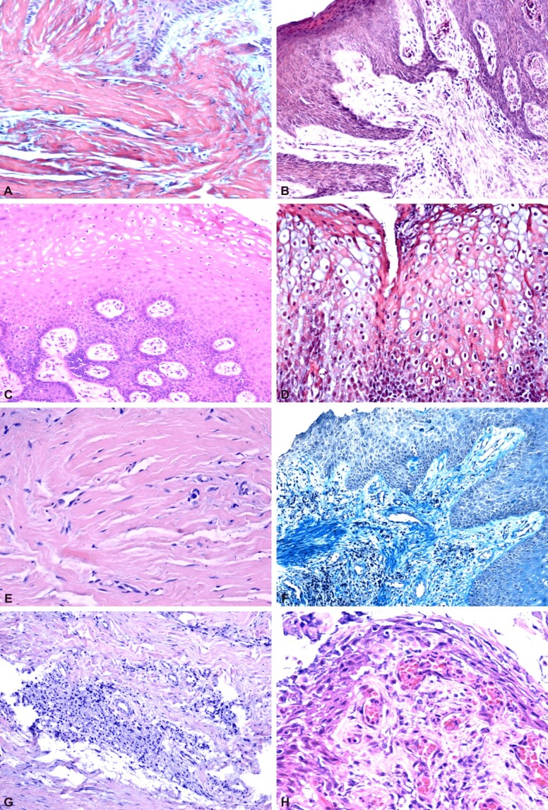
Figure 1.
Peri-implant mucosa: (A) Orthokeratinized epithelium; (B) Parakeratinized epithelium; (C) Acanthosis, deep epithelial ridges, edema of the epithelial cells in the intermediate layer, which, at the level of the superficial layer, have a vacuolar appearance; (D) Edema of cells in all layers of the epithelium; (E) Lamina propria rich in collagen fibers associated with numerous fibroblasts; (F) Diffuse lymphoplasmocytic inflammatory infiltrate; (G) Nodular-looking lymphoplasmocytic inflammatory infiltrate; (H) Numerous capillaries and inflammatory infiltrate in a papilla of the connective tissue. HE staining: (A–C, E and H) ×200; (G) ×100. Masson’s trichrome staining: (D and F) ×100

