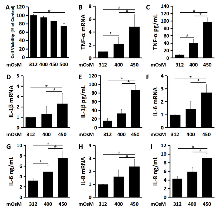
Figure 1.
Hyperosmolarity stimulates inflammatory mediators in primary cultured human corneal epithelial cells (HCECs). (A) Cell viability of HCECs exposed to different osmolarities analyzed by MTT assay. The pro-inflammatory cytokines, TNF-α (B,C), IL-1β (D,E) and IL-6 (F,G), as well as chemokine IL-8 (H,I) significantly increased at mRNA and protein levels in HCECs exposed to a hyperosmolar medium at 400 and 450 mOsM for 4 and 24 h, respectively, compared with isosmolar control at 312 mOsM as evaluated by RT-qPCR and ELISA. Data are presented as mean ± SD of 3 independent experiments. * p < 0.05, compared between two groups.

