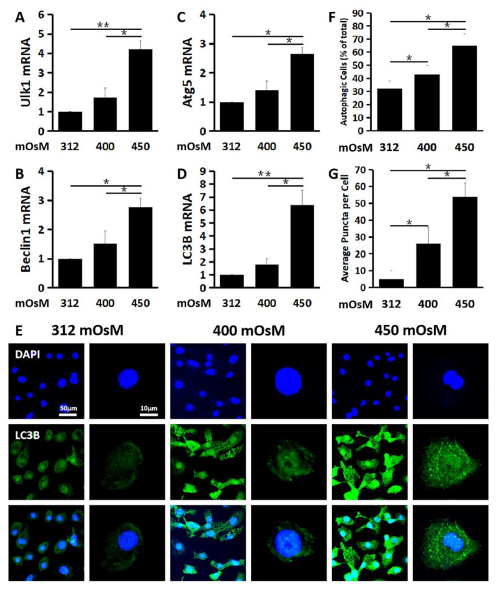
Figure 2.
Hyperosmolarity induced autophagosome formation in primary HCECs. The mRNA expression of Ulk1 (A), Beclin1 (B), Atg5 (C) and LC3B (D) was significantly induced in HCECs exposed to hyperosmolar medium at 450 mOsM for 24 h, as evaluated by RT-qPCR. (E) Representative immunofluorescent images showing the LC3B positive cells with punctate staining in HCECs at various osmotic conditions. (F) Percentage of LC3B positive autophagic cells in HCECs at different osmolarities, evaluated by randomly selected fields with at least 100 cells per sample. (G) Average puncta per autophagic cell. The data are presented as mean ± SD from three independent experiments. * p < 0.05, ** p < 0.01, compared between two groups.

