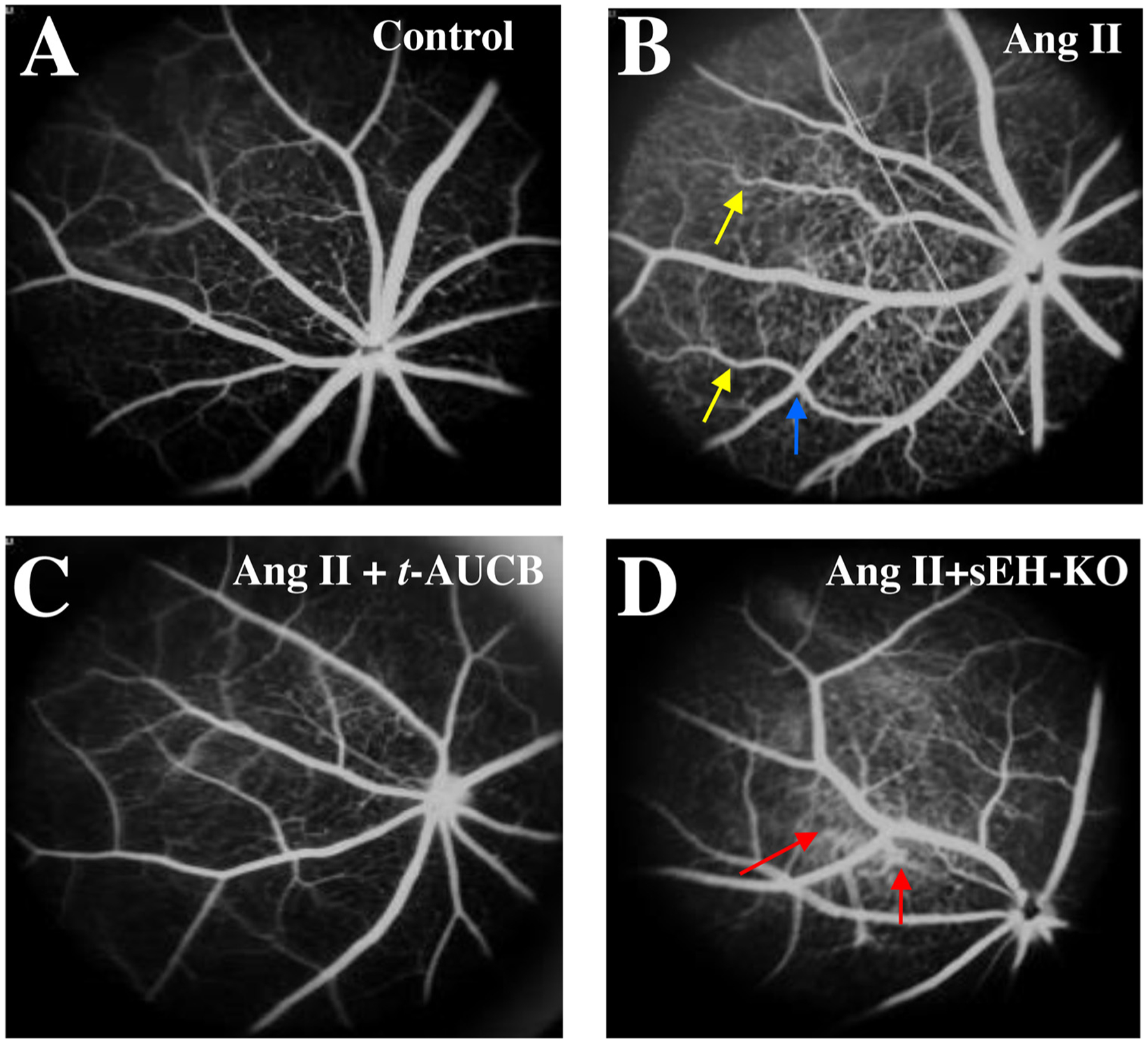
Fig. 5.

Fluorescein angiograms (FA) of the retina in (A) sEH (+/+) + vehicle, (B) sEH (+/+) + Ang II, (C) sEH (+/+) + Ang II + t-AUCB, and (D) sEH (−/−) + Ang II mice. We treated mice given different treatments for four weeks. There were some signs of hypertensive retinal vascular remodeling, such as right-angle crossing (blue arrow) and increased vascular tortuosity (yellow arrow) in the Ang II group. There was marked hyperfluorescence (red arrow) in the sEH (−/−) + Ang II group, suggesting retinal vascular leakage (D).
