
Figure 5. Stroke-induced astrogliosis is prominent at astrocyte endfeet in rat cortex.
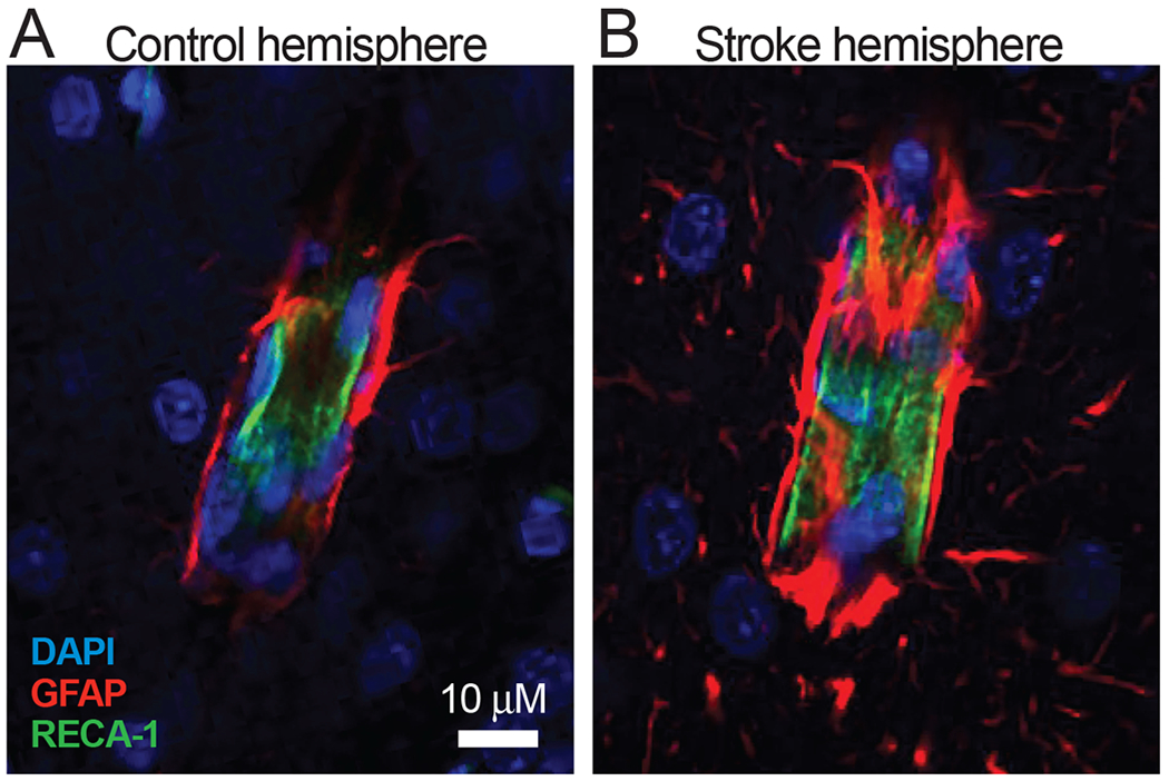
A: GFAP immunolabeled astrocyte endfeet (red) terminating on a cortical vessel in the control hemisphere of a rat that underwent transient experimental stroke. B: Astrocyte endfeet terminating on a vessel in the peri-infarct intact cortical tissue of the stroke hemisphere. Note the increased expression of GFAP and hypertrophy of the endfeet processes, suggesting astrogliosis. DAPI is shown in blue and the rat endothelial cell marker (RECA-1) is shown in green. Images were obtained on a Zeiss LSM 780 (Objective Plan-Apochromat 63x/1.40 Oil) 3 days after middle cerebral artery occlusion in 6 week-old Long Evans rat.