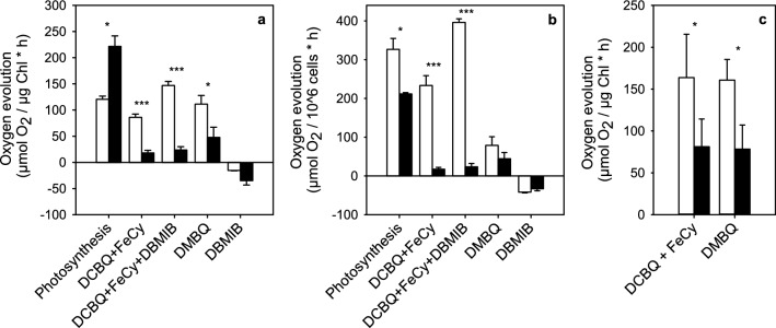
Fig. 4.
PSII activity, measured as light-saturated oxygen evolution from control (white bars) and EL samples of C. reinhardtii (black bars) both in vivo (a, b) and in isolated thylakoids (c) with different electron acceptors and one inhibitor of electron transfer, normalized to Chl (a, c) or cell (b) concentrations. Light-saturated oxygen evolution was measured at PPFD 2000 µmol m−2 s−1 from cultures with OD730 of 0.5. The cultures were grown in PPFD of either 100 or 3000 µmol m−2 s−1 and 1 ml samples were used in measurement. Isolated thylakoids were used in final chlorophyll concentration of 5 µg ml−1. The concentrations of artificial electron acceptors (DCBQ, FeCy and DMBQ) were 0.5 mM and the inhibitor (DBMIB) was added at the concentration of 0.5 µM. Each bar represents an average of three biological replicates and the error bars show SD (*P < 0.05, ***P < 0.005)

