
Figure 5.
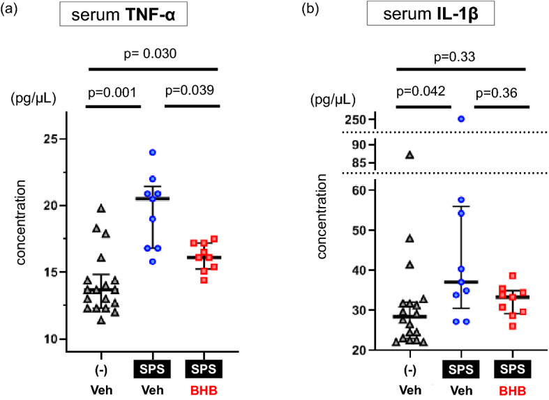
BHB reduces SPS-induced increase in serum inflammatory cytokine levels. (a) Serum TNF-α levels are shown (median [IQR]; non-SPS + Veh: 13.7 [12.4–14.4], SPS + Veh: 20.5 [16.8–20.9], SPS + BHB: 16.1 [15.4–17.2], Kruskal–Wallis test; X2 = 17.265, df = 2, p = 0.0002). SPS exposure increased levels of TNF-α in the blood (Mann–Whitney’s U-test; W = 11, p = 0.001). Chronic BHB treatment decreased levels of TNF-α in the blood in SPS-induced PTSD rats (Mann–Whitney’s U-test; W = 69, p = 0.039). (b) Serum IL-1β levels are shown (median [IQR]; non-SPS + Veh: 28.5 [23.5–31.8], SPS + Veh: 37.1 [33.9–54.3], SPS + BHB: 33.4 [29.7–34.5], Kruskal–Wallis test; X2 = 7.503, df = 2, p = 0.024). SPS increased levels of blood IL-1β (Mann–Whitney’s U-test; W = 34, p = 0.042). Chronic BHB treatment did not significantly decreased levels of IL-1β in the blood of rats exposed to SPS (Mann–Whitney’s U-test; W = 49.5, p = 0.33). The data are presented as scatter plots with median and interquartile range. Kruskal–Wallis test and Mann–Whitney U tests were performed to compare each group. p-values were corrected by Bonferroni method. TNF-α: Tumor necrosis factor-alpha, IL-1β: Interleukin-1 beta, SPS: single prolonged stress, Veh: Vehicle (PBS), BHB: beta-hydroxybutyrate, n = 18 (non SPS + Veh group) or 9 (SPS + Veh group and SPS + BHB group).