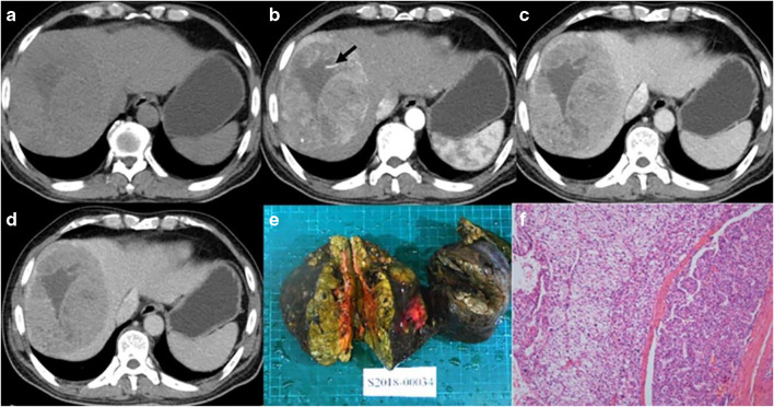
Fig. 5.
Male, 52 years old, HCC with MVI. Panels a–d correspond to the pre-contrast, arterial, portal, and equilibrium phases of CT images, respectively. The tumor with non-smooth margin on axial imaging, and intratumoral arteries can be seen on arterial phase (arrow). Panels e and f are gross specimens of tumor and photomicrograph (H&E, × 100)

