Figure 3.
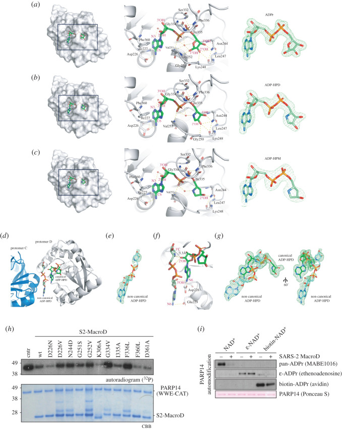
Identification of key residues for ligand binding, catalysis and drug design. (a) Left panel: Surface representation of S2-Macro:ADPr complex showing the tight coordination of ADPr (green) within a deep binding cleft. Middle panel: Ribbon-liquorice representation of ADPr coordination. Residues (black labels) and ADPr atoms (red labels) involved in the interactions are highlighted. Structural waters are given as red spheres and polar interactions as dotted lines. Right panel: Composite omit map (green) of the ADPr ligand contoured at 2σ. (b) As (a) for the S2-MacroD:ADP-HPD complex. (c) As (a) for the S2-MacroD:ADP-HPM complex. (d) Ribbon representation of the S2-MacroD:ADP-HPD complex. An extra ADP-HPD ligand (non-canonical, light green) is visible between chain C (blue) and chain D (white). The canonical ADP-HPD is shown in dark green sticks. (e) Composite omit map of the second bound ADP-HPD ligand contoured at 2σ. (f) Ribbon-liquorice representation of the interaction between the two ADP-HPD ligands within protomer D. Polar protein-ligand and ligand–ligand interactions are shown in dashes lines (yellow). Measured distance (3.3 Å) between the C1″ atom of the non-canonical ligand and the 2′OH moiety of the canonical ligand is shown as black line. Ligand atoms are labelled in red and S2-MacroD residues in black. (g) Composite omit map of both ligands contoured at 2σ and shown in two different orientations. (h) S2-MacroD variants carrying amino acid substitutions were analysed for their effect on (ADP-ribosyl)hydrolase activity. The model substrate PARP14 WWE-CAT was modified to completion in presence of 32P-NAD+ and subsequently incubated with either wt or S2-MacroD mutants as indicated. Samples were analysed by CBB staining and autoradiography. (i) Substitutions at the adenosine base affect S2-MacroD (ADP-ribosyl)hydrolase activity. The model substrate PARP14 WWE-CAT was modified in the presence of either β-NAD+, biotin-NAD+ or ε-NAD+ as indicated. The reactions were subsequently incubated in the absence or presence of S2-MacroD as indicated. Samples were blotted onto a nitrocellulose membrane and analysed by Ponceau S staining and substitution specific detection reagents.

