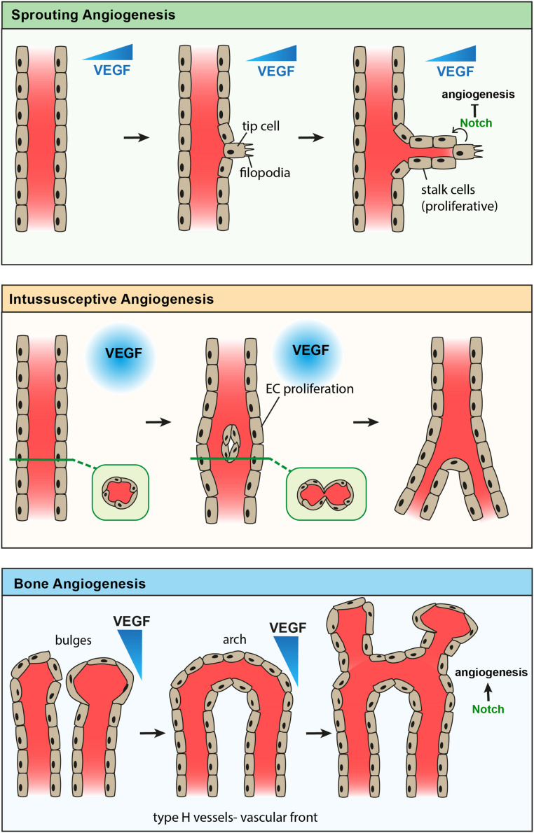
FIGURE 1.
Comparison of known modes of angiogenesis. Sprouting angiogenesis (top) showing formation of an endothelial tip cell with filopodia in response to a gradient of VEGF. Stalk cells proliferate to elongate the new vessel branch, while Notch signaling from the tip cell inhibits further tip cell formation. Intussusceptive angiogenesis (middle) showing endothelial cell proliferation in response to high levels of VEGF, leading to the formation of a new endothelial vessel wall which splits one vessel into two. Angiogenesis of type-H vessels in bone (bottom) showing anastomosis of bulge structures, forming an arch structure. Endothelial cells proliferate to form new bulge structures in the direction of a VEGF gradient, while Notch signaling promotes angiogenesis in bone.

