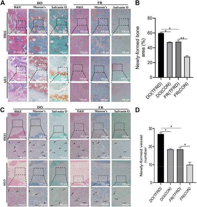
FIGURE 3.
TFRD accelerates bone healing by promoting bone and vessel formation in rats of DO and FR models. (A) Representative histological images and (B) quantification of newly-formed bone at 45 days after surgery (n = 3 per group). (C) Representative histological images and (D) quantification of newly-formed vessels at 17 days after surgery (5 random visual fields per section, three sections per staining, nine sections per rat and three rats per time point). From left to right: H&E, Masson’s and Safranin O staining. The black boxes represent higher-magnification view. The data are expressed as the mean ± SEM of three independent experiments; Black arrows indicate micro-vessels. *p < 0.05, **p < 0.01, NS: not significant.

