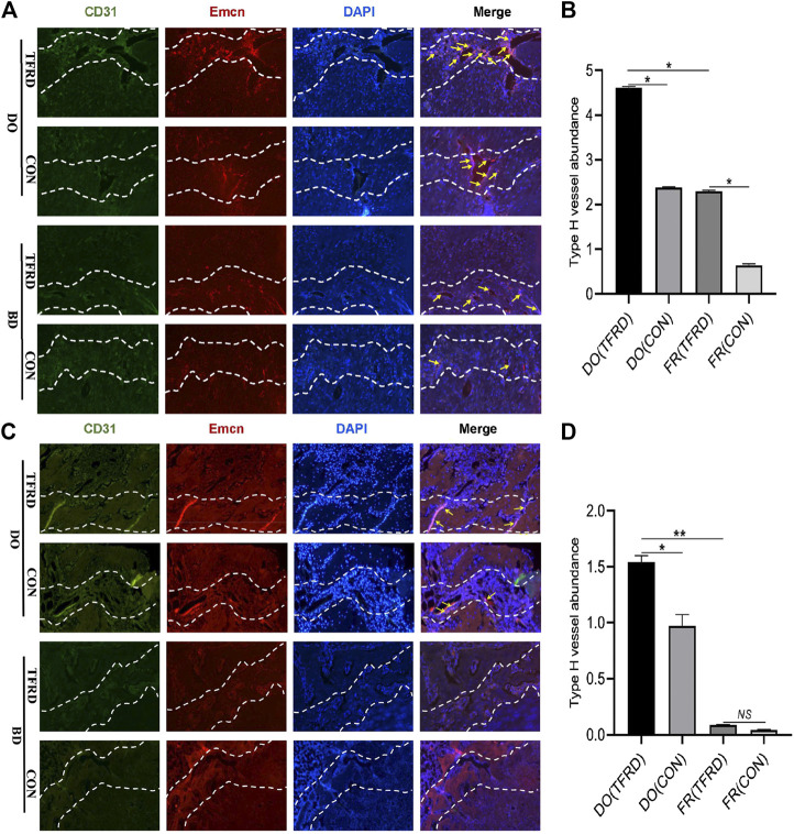
FIGURE 4.
TFRD increases the abundance of type H vessels during bone healing process. (A) Representative immunostaining images of CD31 (green), Emcn (red) and Nuclei (blue, stained with DAPI) inside bone fracture or distraction regions of four groups at 17 and (C) 45 days after surgery (n = 3 per time point). The region between the two white dotted lines represents the bony gaps including bone fracture or distraction zone. The distraction zone was caused by gradual and controlled tensile stress. The type H vessels were marked with yellow arrows. (B), (D) Quantification of the abundance of CD31+Emcn+ (type H) vessels (light pink) at 17 and 45 days after surgery, respectively. The abundance of type H vessels is represented by the percentage of CD31+Emcn+ vessel area inside the bony gap. The data are expressed as the mean ± SEM of three independent experiments, *p < 0.05, NS: not significant.

