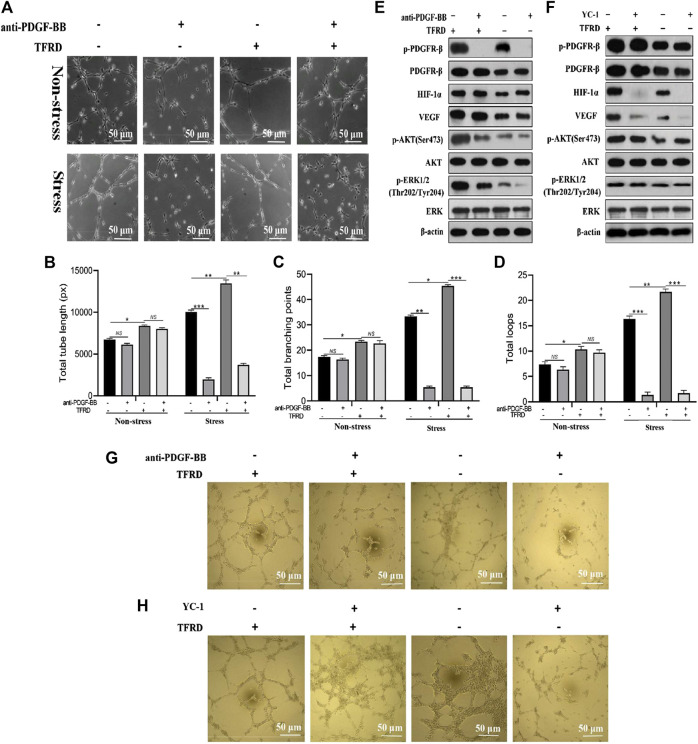
FIGURE 7.
PDGF-BB/PDGFR-β instead of HIF-1α/VEGF axis involves in the TFRD-promoted angiogenesis under stress conditions. (A) Representative images of tube formation of EPCs stimulated with 100 μg/ml TFRD or 20 μg/ml function blocking anti-PDGF-BB antibody for 6 h under stress or non-stress conditions. (B) Quantification of total tube length, (C) total branching points and (D) total loops of EPCs in five randomly chosen fields. (E) Protein levels of p-PDGFR-β, HIF-1α, VEGF, p-PI3K and p-ERK1/2 in the stretched EPCs treated with or without TFRD for 6 h after the stimulation with 20 μg/ml function blocking anti-PDGF-BB antibody or (F) 10 μM HIF-1α inhibitor (YC-1) for 1 h. (G) Representative images of tube formation of the stretched EPCs stimulated with or without TFRD for 6 h after the stimulation with 20 μg/ml function blocking anti-PDGF-BB antibody or (H) 10 μM HIF-1α inhibitor (YC-1) for 1 h. The data are expressed as the mean ± SEM and all experiments were performed at least three times. *p < 0.05, **p < 0.01, ***p < 0.001, NS: not significant.

