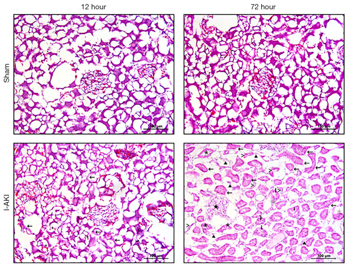
Figure 2.
Histopathological changes of dog kidneys with ischemic acute kidney injury (I-AKI). The kidney samples were collected from the sham and ischemia/reperfusion (I/R) groups at 12 and 72 h after I/R operation, and stained using hematoxylin and eosin (H&E) method. >, atrophic renal tubules; *, exfoliated cells; ▲, fibrotic tissue; ↓, protein cast; ★, glomerular atrophy; ←, cell swelling, proliferation, lumen stenosis, and brush-like edge disappearance.

