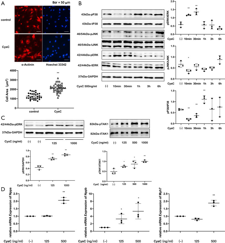
Figure 6.
Effects of CysC on the development of myocardial hypertrophy in rat primary cardiomyocytes. (A) Immunofluorescence staining of α-actinin in primary cardiomyocytes and measurement of cross-sectional area; (B) MAPK phosphorylation of primary cardiomyocytes after intervention with CysC (500 ng/mL) at different time points; (C) ERK and TAK1 phosphorylation of primary cardiomyocytes after 10-minute intervention with CysC of different concentrations; (D) gene expression of Nppa, Nppb, and Myh7 of primary cardiomyocytes after 6-hour intervention with CysC of different concentrations (*, P<0.05; **, P<0.01). ERK, extracellular regulated protein kinase; JNK, c-Jun N-terminal kinase; TAK1, transforming growth factor activated kinase-1; GAPDH, glyceraldehyde-3-phosphate dehydrogenase; CysC, cystatin C.

