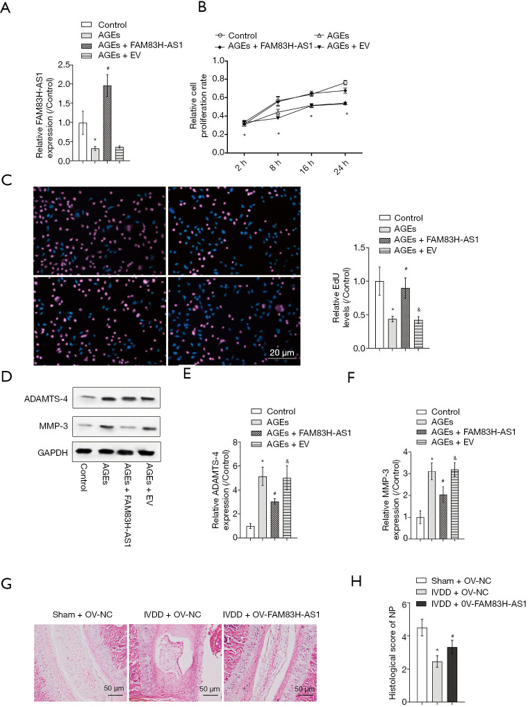
Figure 2.
Overexpression of FAM83H-AS1 promoted NP cell proliferation, alleviated ECM degradation and prevent IVDD progression in vivo. NP cells isolated from IVD tissues were treated with 50 µg/mL AGEs, followed by transfection with FAM83H-AS1 plasmid or empty vector. NP cells treated with PBS served as the control. (A) The efficiency of FAM83H-AS1 overexpression was verified by qRT-PCR analysis; (B) CCK-8 assay was performed to assess the viability of NP cells; (C) cell proliferation capacity was also determined using the EdU incorporation assay; (D,E,F) the protein expression of ADAMTS-4 and MMP-3 was measured by western blot. (G) Representative H&E staining of disc samples from different experimental groups at 4 weeks post-surgery. (H) Histological scores at 4 weeks post-surgery in three groups. All experimental data are shown as mean ± SD from at least 3 independent assays. *, P<0.05 vs. control group; #, P<0.05 vs. AGEs group; &, P<0.05 vs. IVDD + FAM83H-AS1 group. ECM, extracellular matrix; NP, nucleus pulposus; AGEs, advanced glycation end products; qRT-PCR, quantitative real-time PCR.

