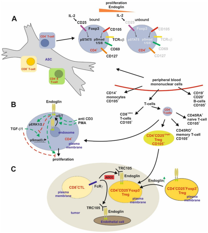
Figure 6.
Endoglin expression and function in T cells. (A) Adipose tissue derived stem cells (ASC) are able to regulate T-cell dependent immune responses in part by sequestering reactive CD4+ T-cells. Allo-activated CD4+ T cells tightly bound to ASC show a higher CD25 expression and in turn IL-2-dependent STAT5 activation. These cells present a higher proliferation and increased Endoglin (Eng) expression along with a higher Smad1/5/8 phosphorylation (activation). Nevertheless, aside the higher Foxp3 expression, the bound cells also had a higher CD127 expression excluding that they are Tregs. (B) High expression of Eng is present in CD4+ T-cells isolated from peripheral blood. TCR/pathway activation of these CD4+ T-cells by CD3 or PMA leads to a higher surface expression of Eng and increased serine/threonine phosphorylation of the receptor. Activation of Eng induces ERK1/2 phosphorylation and higher proliferation. On the other hand, Eng interferes with TGF-β1-mediated Smad signaling and inhibition of proliferation. (C) In the setting of tumorigenesis, intratumoral CD4+ T-cells, i.e., CD25+/Foxp3+ Tregs, express higher Eng compared to their circulating counterparts. This causes an antibody-dependent cellular cytotoxicity (ADCC) resulting in reduction of intratumoral Tregs (immunosuppressive) upon application of TRC105 via CD8+ T-cells.

