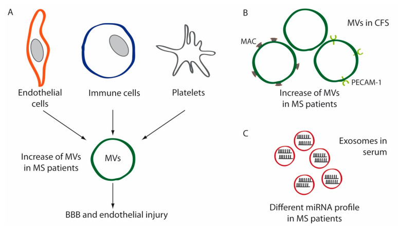
Figure 2.
Schematic diagrams depicting the implication of extracellular vesicles (EVs) in MS. (A) MVs secreted by endothelial cells, immune cells and platelets are increased in the plasma of MS patients compared to healthy controls. These MVs contribute to blood–brain barrier (BBB) disruption and endothelial injury in these patients. (B) MVs isolated from the cerebrospinal fluid (CSF) of MS patients are increased compared to controls. These EVs are enriched in membrane attack complex (MAC) components and platelet-endothelial cell adhesion molecule-1 (PECAM-1). (C) Circulating exosomes have a different miRNA profile in MS patients.

