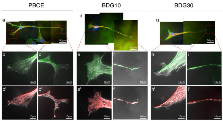
Figure 4.
Coordinate nucleation of F-actin and Microtubules drives the hBM-MSCs’ shape on PBCE, BDG10 and BDG30. (a,d,g) Representative fluorescence images of hBM-MSC in proliferation medium in the absence of FBS (F-actin [FITC-phalloidin, green], Microtubules [human anti-tubulin antibody, red], and nuclei [DAPI, blue]). Due to the length of the cells, the images include 2/3 microscope frames. Elaboration of selected details ((b,c,bI,cI); (e,f,eI,fI); (h,i,hI,iI)) revealed the coordinate action of F-actin and tubulin in building the new cytoskeleton architecture of the cell as a consequence of the seeding on polymer films.

