Figure 5.
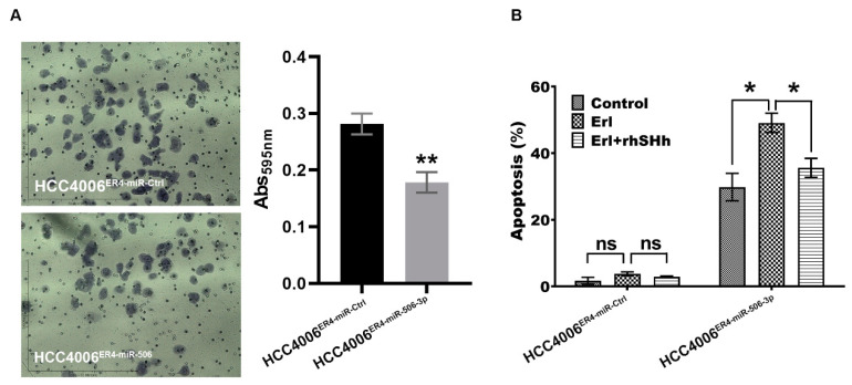
Migration and apoptotic activity of erlotinib-resistant cells and miR-506-3p-overexpressing erlotinib-resistant cells. (A) The migratory potential of HCC4006ER4-miR-Ctrl and HCC4006ER4-miR-506-3pOE cells was determined by the transwell migration assay. The photomicrographs represent the difference in the in vitro migration of HCC4006ER4-miR-Ctrl and HCC4006ER4-miR-506-3pOE cells toward the serum for 24 h (Magnification, 10×). Histogram showing crystal violet absorbance at 595 nm. Values in the bar graphs represent the mean ± SD (n = 6). ** p < 0.001 compared to HCC4006ER-miR-Ctrl cells. (B) Overexpression of miR-506-3p vectors in HCC4006ER4 cells promoted apoptosis after exposure to erlotinib which is counteracted by treatment with both erlotinib and rhSHH protein. Data are expressed as the mean ± SD, ** p < 0.001.

