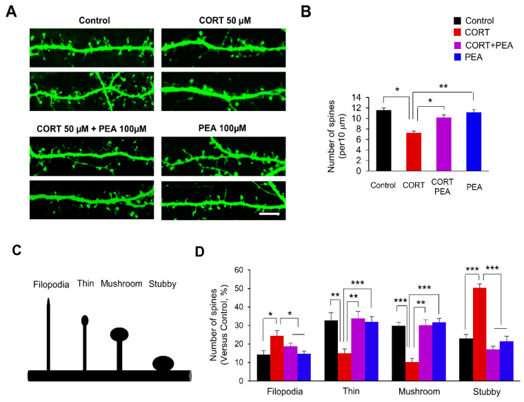
Figure 1.
Treatment with 2-Phenethylamine (PEA) restores dendritic spine formation in corticosterone (CORT)-induced hippocampal neurons. (A) PEA restores dendritic spine density in CORT-induced hippocampal neurons. Cultured hippocampal neurons were isolated from E18 embryos, cultured and transfected with a GFP plasmid at DIV 10. After 4 days, neuronal cultures were administrated with the CORT (50 μM) for 24 h followed by treatment with PEA (100 μM) for 24 h. Scale bar, 10 μm. (B) Quantification of the number of dendritic spines in each condition. n = 15 neurons from three independent cultures using three mice for each condition. Statistical significance was determined by two-way ANOVA with Bonferroni correction test. Data are shown as relative changes versus controls. * p < 0.05, ** p < 0.01 (C) Different types of dendritic spines (Filopodia, thin, mushroom, and stubby). (D) PEA restores dendritic spine morphology in CORT-induced hippocampal neurons. n = 10 cultured cortical neurons and 300 dendritic spines for each condition. Statistical significance was determined by two-way ANOVA with Bonferroni correction test. Data are shown as relative changes versus controls. * p < 0.05, ** p < 0.01, *** p < 0.001.

