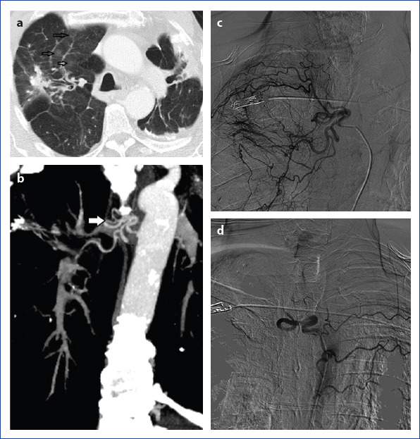
Figure 1.

Patient with hemoptysis due to bronchiectasis. Axial section CT image (a) shows bilaterally bronchiectasis, sequels and right upper lobe focal ground-glass opacity because of alveolar hemorrhage. Coronal reconstruction section CT image (b) shows the right abnormal bronchial vessels. In angiography (c), dilatation, hypertrophy and abnormal vascularity at the intercostal branch and bronchial artery originating from right intercostobrachial truncus. Totally obstruction at the right intercostobrachial truncus after PVA embolization (d). Left intercostal branches are visible due to the reflux of contrast.
