
Figure 3.
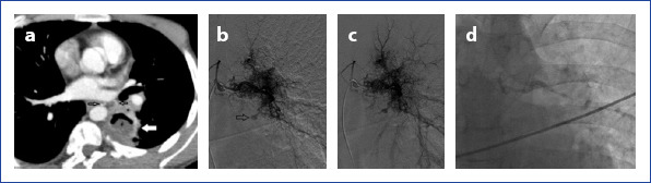
Axial CT image shows left lower lobe cavitary lesion compatible with a thick wall abscess. An abnormal dilated bronchial artery adjacent to the cavity. In angiographic images (b) demonstrate right and left bronchial arteries originate from the same truncus. Dense pathological vascular system arising from the left bronchial artery is obvious. Left bronchial artery is markedly tortuous. A small aneurysmatic enlargement exists in this pathologic region. Fistulization at late images (c) was seen between the bronchial artery and pulmonary vascular system. In angiography (d) abnormal left bronchial artery occlusion after histoacryl embolization was observed.