Figure 4.

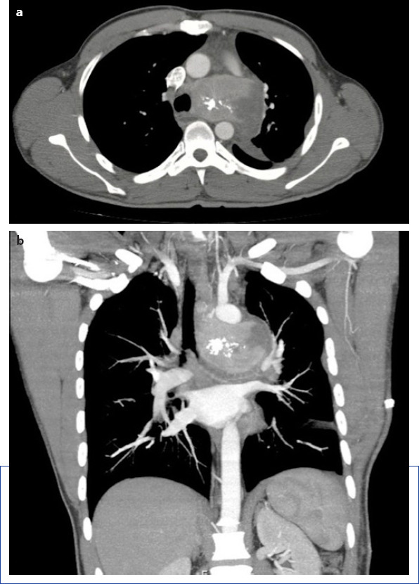
In the axial section of CT examination (a) shows nonhomogenous calcified mass filling aortapulmonary window. The coronal section of CT examination (b) demonstrates mass compression to the adjacent structures.

In the axial section of CT examination (a) shows nonhomogenous calcified mass filling aortapulmonary window. The coronal section of CT examination (b) demonstrates mass compression to the adjacent structures.