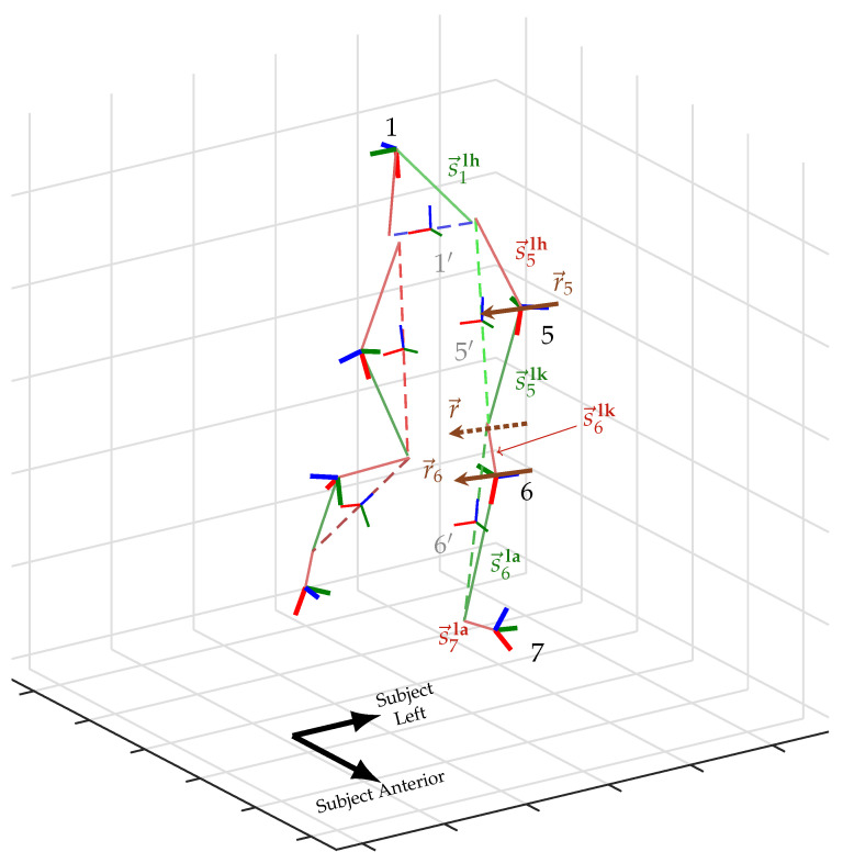
Figure 1.
The human-IMU kinematic system, with the subject mid-stride. Image is an excerpt frame from a 3D animation using the proposed method. The subject’s left leg is labeled with coordinate systems of the IMUs (bold RGB triplets with black text label) and anatomical segments (thin RGB triplets with gray text label), the static vectors from the IMUs to neighboring joint centers (red and green), and the knee’s hinge axis (dotted brown) with their static representations in the thigh and shank IMU frames (solid brown). Notation of variables is detailed in Section 2.1.

