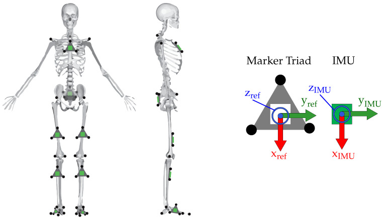
Figure 4.
(Left) Placement of the reflective markers (black circles) and IMUs (green squares) on the subject. IMUs on the thigh and shank were not placed precisely, and location varied both vertically and in the transverse plane. (Right) A blown-up illustration of the marker triads with three markers affixed and IMU. Coordinate system of the IMU was known a priori, and the comparison reference coordinate system of the marker triad was constructed to match.

