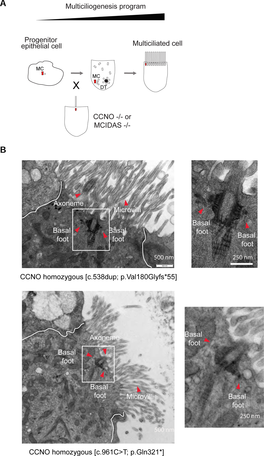
Figure 5. Hybrid cilium formation is independent from other motile cilia in airway multiciliated cells.

(A) Cartoon depicting a simplified version of the multiciliogenesis cellular program. Note CCNO and MCIDAS loss of function mutations lead to oligocilia phenotype. MC, mother centriole. DT, deuterostome. (B) Left: TEM micrographs of a human airway multiciliated cells isolated from nasal epithelium of PCD patients with CCNO loss of function early termination mutations. Boxed areas represent basal bodies with multiple basal feet. Red arrowheads indicate axoneme, basal feet and microvilli. Right: High-magnification views of boxed area. Scale bars represent 500 nm (left) and 250 nm (right).
