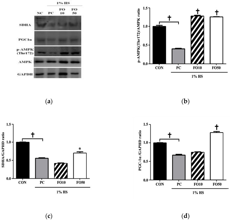
Figure 7.
(a) image of Western blot analysis. Changes in (b) phospho-adenosine monophosphate-activated protein kinase (p-AMPK), (c) succinate dehydrogenase (SDHA) and (d) peroxisome proliferator-activated receptor gamma coactivator-1 α (PGC-1α levels were determined in C2C12 muscle cells following administration of 10 and 50 μg/mL of FO. Each bar represents the mean ± SD. A one-way ANOVA test was carried out to determine significant differences (* p < 0.05 vs. positive control, † p < 0.001 vs. positive control).

