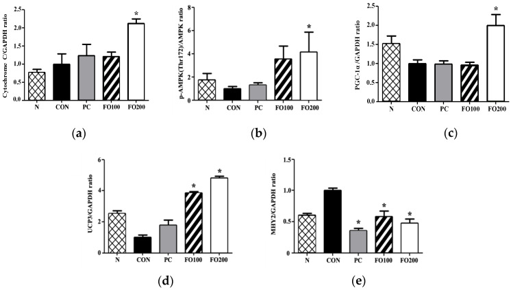
Figure 11.
Immunohistochemical analysis of (a) cytochrome C, (b) p-AMPK (Thr172), (c) PGC-1a, (d) UCP3 and (e) MYH2 was carried out to measure changes following administration of 100 and 200 μg/mL of FO. Each bar represents the mean ± SD. A one-way ANOVA test was carried out to determine significant differences (* p < 0.05 vs. Control).

