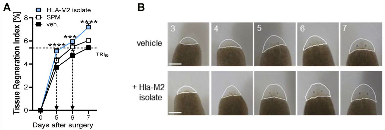
Figure 6. Enhancement of Planarian Regeneration by Exposure to Hla-Treated M2 Macrophage Isolates.

(A) Tissue regeneration index of planarians treated with Hla-M2 isolates, SPMs (200 nM RvD5, RvD2, MaR1, PD1, and 18-HEPE), or vehicle (0.5% MeOH) after the indicated days. Results are means + SEM; n = 14–15 planarians. ***p < 0.001, ****p < 0.0001 Hla-M2 isolate versus vehicle; #p < 0.05 SPMs versus vehicle; one-way ANOVA with Tukey’s multiple comparisons test.
(B) Planarians were treated with LM isolates obtained from M2s that were treated with 1 μg/mL Hla (Hla-M2 isolates) or with 0.5% MeOH (vehicle). Images shown are representative planarians from n = 15 taken at the indicated time points. The outline of the quantified area in each of the images is shown.
