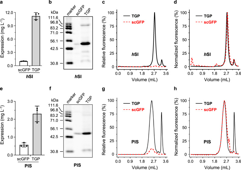
Fig. 6. Replacing GFP with TGP improved expression of two human membrane proteins in mammalian cells.
Assessment of hSI expression was based on fluorescence counts (a), in-gel fluorescence (b), and relative FSEC intensity (c). Normalized FSEC traces of hSI-TGP and hSI-scGFP are shown in d. Assessment of PIS expression was based on fluorescence counts (e), in-gel fluorescence (f), and relative FSEC intensity (g). Normalized FSEC traces of PIS-TGP and PIS-scGFP are shown in h. Mean and standard deviation (a, e) or a representative (b–d, f–h) of three independent experiments on cells with different passage numbers are shown. In c, d, g, and h, TGP traces are shown as black solid lines and scGFP traces are shown as red dashed lines. FSEC fluorescence-detection size exclusion chromatography, GFP green fluorescent protein, TGP thermostable GFP.

