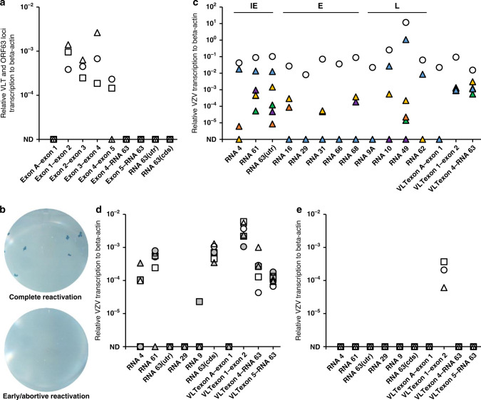
Fig. 4. Effect of anisomycin treatment on VZV transcription in latently VZV-infected sensory neurons in vitro.
a RT-qPCR analysis for transcription across VLT and ORF63 loci in latently VZV-infected human iPSC-derived sensory neurons (HSN) cultures (n = 3). Data on individual HSN culture experiments are shown as unique symbols. b, c Latently VZV-infected HSN cultures were depleted of neurotrophic factors (NGF [nerve growth factor], GDNF [glial-derived neurotrophic factor], BDNF [brain derived neurotrophic factor] and NT-3 [neurotrophin-3]) and treated with anti-NGF antibody (Ab) for 14 days. In total, n = 40 independent cultures were subjected to reactivation stimuli. b Representative examples of a HSN cultures showing complete reactivation (2/40) and early/abortive reactivation (38/40) by infectious focus forming assay after transferring HSN onto ARPE-19 cells. c Representative examples of viral gene expression in cultures showing complete reactivation (n = 1 shown; white circle) and early/abortive reactivation (n = 5 shown; colored triangles). d, e HSN cultures were treated with d anisomycin (n = 6), or e DMSO as solvent control (n = 3) at both the somal and axonal compartment for 1 h, washed twice and cultured for 7 days before RT-qPCR analysis. Data on individual HSN culture experiments are shown as unique symbols and/or colors. Only VLT exon 1–2 is shown as representative of VLT. Source data are provided as a Source Data file (a–e).

