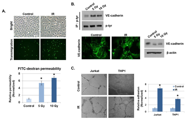
Figure 2.
IR enhances vascular barrier permeability and leukocyte adhesion. (A) Representative images show the morphology of human umbilical vein endothelial cells (HUVECs) and transmigration of THP1 cells co-cultured with HUVECs 24 h after IR. The graph shows the amount of FITC–dextran leakage. Results were normalized to those in control cells (mean ± SD); * p < 0.01. (B) Twenty-four hours after IR, immunofluorescence staining for vascular endothelial (VE)–cadherin in IR-treated HUVECs was performed. VE–cadherin and phosphorylated VE–cadherin levels were measured by Western blotting. β-actin and p-tyr were used as protein loading controls. (C) HUVECs were seeded onto Matrigel, subjected to IR (5 Gy), and co-cultured with leukocytes (THP1 and Jurkat cells) for 6 h. The graph shows the relative adhesion rate of leukocytes to HUVECs (mean ± SD); * p < 0.01.

