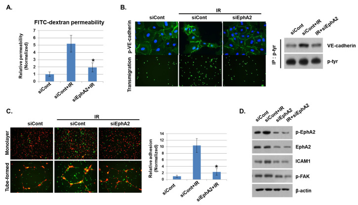
Figure 4.
Depletion of EphA2 with siRNAs blocks IR-induced endothelial cell damage. (A) HUVECs were transfected for 24 h with EphA2 siRNAs, which was followed by IR (5 Gy) exposure for an additional 24 h. Endothelial permeability is represented by the amount of FITC–dextran staining. * p < 0.05. (B) Twenty-four hours after IR, siCont or siEphA2-treated HUVECs were stained with FITC-conjugated p-VE–cadherin and visualized using immunofluorescence (upper panel). Carboxyfluorescein succinimidyl ester (CFSE)-labeled THP1 cell transmigration when co-cultured with siCont or siEphA2-treated HUVECs (lower panel). Western blotting results show levels of p-VE–cadherin in siCont or siEphA2-treated HUVECs after IR. P-tyr was used as a protein loading control. (C) Monolayers or tubes of PKH26-labeled HUVECs (red) were exposed to IR and then co-cultured with CFSE-labeled THP1 cells (green) for 24 h. The graph shows the relative adhesion rate. * p < 0.05. (D) Expression levels of p-EphA2, EphA2, ICAM1, and p-FAK in HUVECs were determined using Western blotting. β-actin was used as a protein loading control.

