Figure 6.
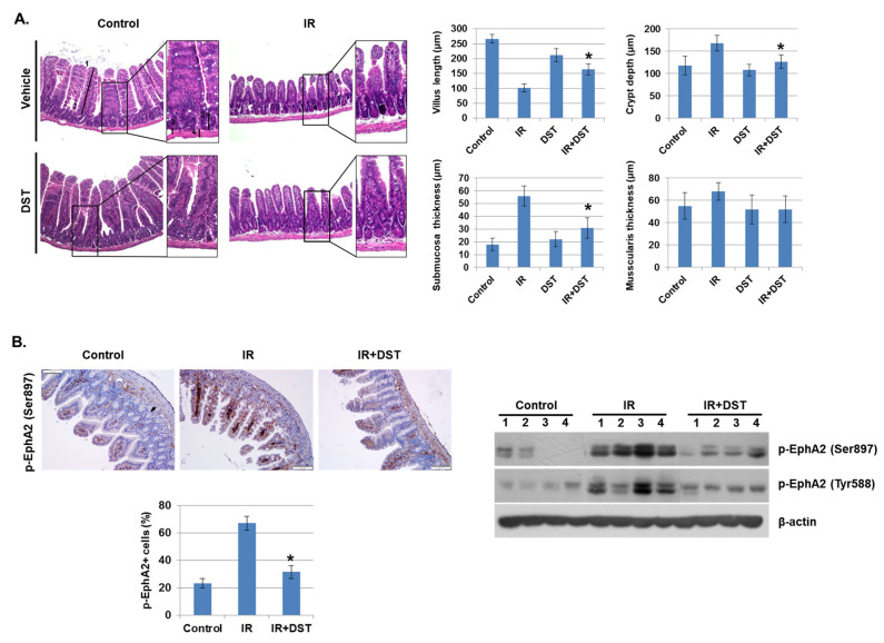
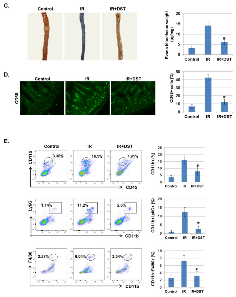
Dasatinib suppresses intestinal injury by radiation exposure in mice. Mice were exposed to IR (5 Gy) followed by treatment with dasatinib. Twenty-seven days after IR, mice were sacrificed. (A) Representative images show hematoxylin and eosin (H&E) staining from intestine of control, IR, dasatinib (DST) and IR+DST animals. The sections were analyzed for villus length (1), crypt depth (2), submucosa thickness (3) and muscularis thickness (4) (original magnification ×40, insert ×100). Each bar represents mean ±SD of 7–9 analyzed sections per animal (n = 5). * p < 0.05. (B) Representative images show EphA2 (Ser897) expression in mouse intestine using immunohistochemical staining. The graph indicates the percentage of p-EphA2-positive cells. Expression of p-EphA2 (Ser897 and Tyr588) was measured in mouse intestinal tissues by Western blotting; n = 4. β-actin was used as a protein loading control.; mean ± SD, * p < 0.05. (C) Intestinal permeability was assessed using the Evans blue assay. Representative images show mouse intestine. The graph shows the measurement of Evans blue dye leakage into the intestine; mean ± SD, * p < 0.05. (D) Immunofluorescence staining of CD68 in mouse intestine. The graph shows the percentage of CD68-positive cells; mean ± SD, * p < 0.05. (E) Mouse intestinal lamina propria cells were stained for CD45/CD11b/Ly6G/F480 and analyzed by flow cytometry. Representative images show CD11b+ (myeloid cells), CD11b+Ly6G+ (neutrophils), and CD11b+F4/80+ (macrophages); mean ± SD, n = 5, * p < 0.05.


