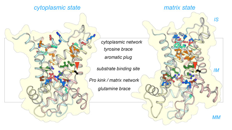
Figure 3.
CpAAC has all of the key features of a functional ADP/ATP. Structural models of CpAAC, in the cytoplasmic state (left) and matrix state (right), based on Protein Data Bank (PDB) entries 4c9g [16] and 6gci [28] respectively, calculated by SWISS-MODEL [38]. Repeats 1, 2, and 3 are colored in pastel blue, yellow, and red, respectively. The residues of the Pro-kink (brown), matrix network (red and blue), glutamine-brace (cyan), substrate binding site (green), aromatic plug (orange), tyrosine-brace (cyan), and cytoplasmic network (red and blue) are indicated. Residue C238 in CpAAC is indicated by a red arrowhead. IS, intermembrane space; IM, mitochondrial inner membrane; MM, mitochondrial matrix.

