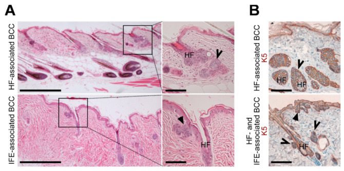
Figure A3.
BCC-like tumors of Ptchf/f K5CreERT skin arise from HF and IFE cells. (A,B) Representative (A) H&E stainings and (B) anti-K5 antibody stainings of BCC from 28-37-week-old Ptchf/f K5CreERT back skin (N = 5). Please note that the K5CreERT-deleter is leaky, and thus, BCC-like lesions also develop without tamoxifen induction [15]. Solid arrows: BCC associated to the IFE and open arrows: BCC associated to HF. HF, hair follicle. Boxes: zoomed areas. Scale bars: 20 µm (A, left) and 100 µm (A, right and B).

