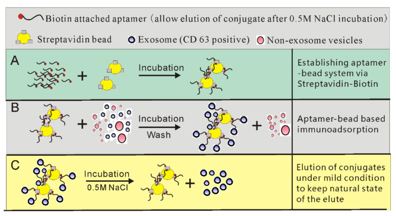
Figure 6.
A schematic of the establishment of a bead/aptamer system for native exosome isolation. (A) Synthesizing 3′ biotinylated CD63-1 aptamer and incubating with streptavidin magnetic beads to prepare the aptamer–magnetic beads system. (B) The aptamer–magnetic beads are incubated with exosome-containing solution. After incubation and washing to remove impurities, exosomes displaying CD63 expression can be isolated. (C) Because the aptamer used in this system undergoes spatial changes after 0.5 M NaCl incubation (mild condition) and releases the conjugate, the natural structure and biological functions of the collected exosomes are ensured.

