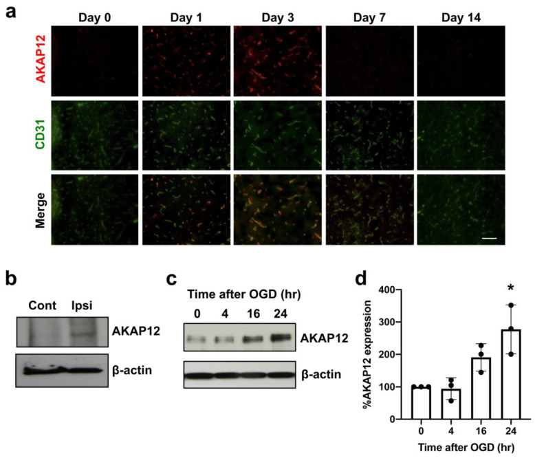
Figure 1.
AKAP12 expression after stroke: (a) Double-staining of AKAP12 (red) with an endothelial marker CD31 (green) showed that AKAP12 was expressed in or around blood vessels, and its expression level transiently increased after stroke. Scale bar = 50 μm. (b) Western blot using endothelial fractions from stroke mice confirmed that compared to the contralateral hemisphere (Cont), AKAP12 level was elevated in the brain endothelium of the ipsilateral hemisphere (Ipsi) at 3 days after stroke. (c,d) In HBMECs, OGD/Reoxygenation stimulation, an in vitro model to mimic in vivo cerebral ischemia-reperfusion, induced an upregulation of AKAP12. Mean ± SD of n = 3. * p < 0.05 vs. control (0 hr after OGD). Kruskal-Wallis test followed by post-hoc Dunn’s multiple comparison test.

