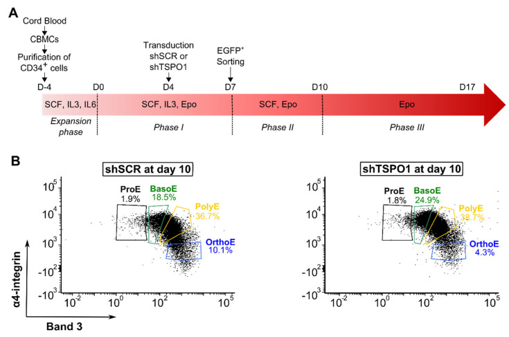
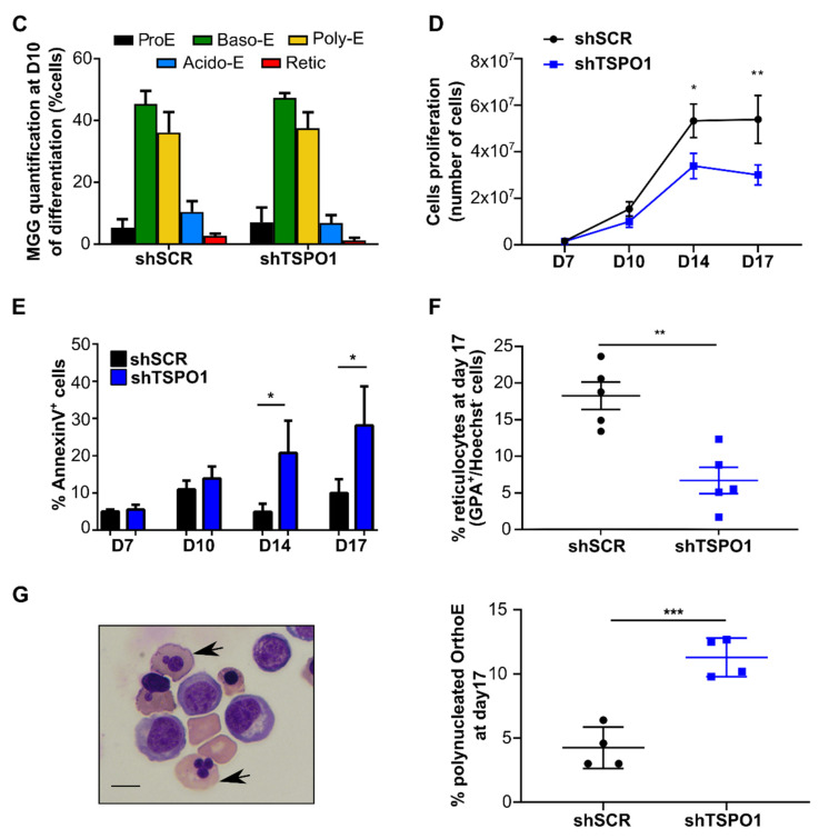
Figure 1.
TSPO1 downregulation does not affect differentiation kinetics but diminished enucleation rate. (A) Schematic of the ex vivo erythroid differentiation protocol following shRNA-mediated downregulation of TSPO1 (CBMC, cord blood mononucleated cells; shSCR, scramble shRNA; shTSPO1, TSPO1 shRNA). CD34+ progenitors were isolated from cord blood and transduced at Day 4 with a lentiviral vector harboring either the TSPO1 shRNA or scramble shRNA, together with the green fluorescent protein (EGFP) transgene. EGFP+ cells were sorted at Day 7 and differentiated until Day 17. (B) Representative profile in flow cytometry of cells at Day 10 of erythropoiesis in scramble shRNA and TSPO1 shRNA. Glycophorin A positive (GPA+) cells are gated according to α4-integrin and Band 3 expression levels to discriminate the different stages of differentiation. (C) May-Grünwald Giemsa (MGG)-based quantification at Day 10 of erythroid differentiation. (D) Erythroblast proliferation at Day 7, 10, 14, and 17 (n = 4). (E) Apoptosis assay (annexin V) by flow cytometry at Day 7, Day 10, Day 14, and Day 17 (n = 3). (F) Enucleated cells (GPA+/Hoechst- cells) were quantified by flow cytometry at Day 17 of differentiation (n = 5). (G) Representative images (left) and quantification (right) of polynucleated Ortho-E (arrow) at Day 17 after MGG coloration (n = 3). Scale bar = 20 μm. * p < 0.05, ** p < 0.01, *** p < 0.001.


