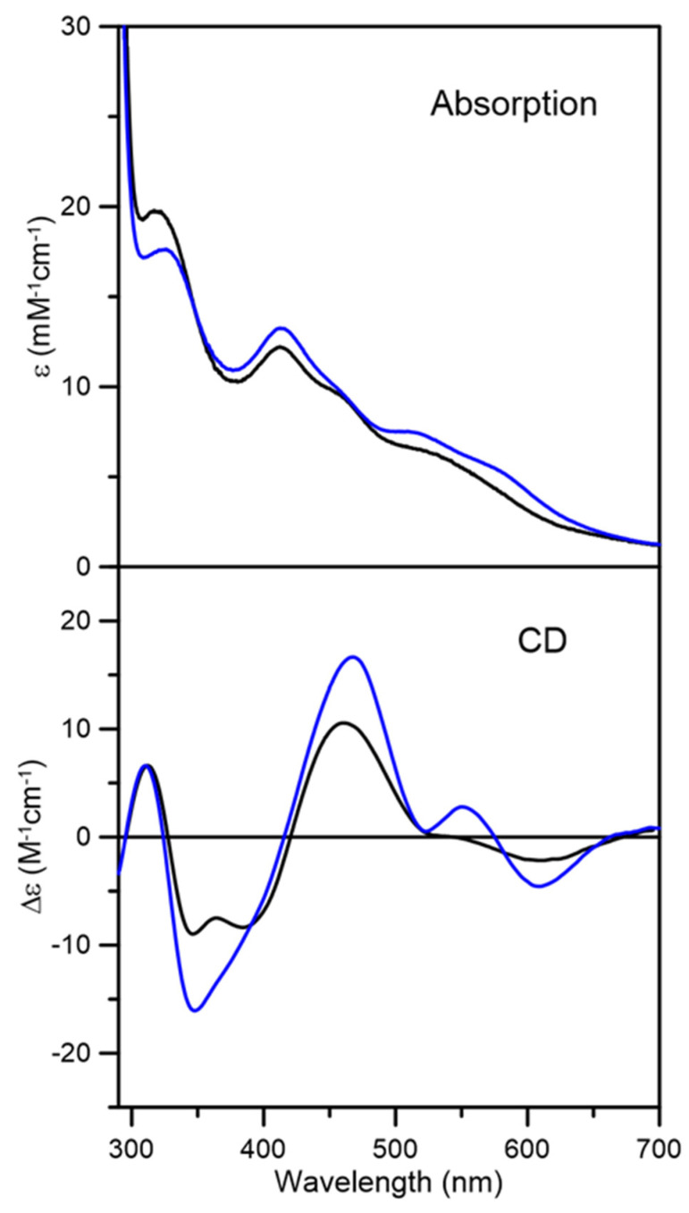
Figure 3.
UV-visible absorption and circular dichroism spectra of reconstituted At GRXS15. Fraction 1 (black line) and fraction 2 (blue line) were obtained after separation using a Mono-Q column. Spectra were recorded under anaerobic conditions in sealed 0.1 cm cuvettes in 100 mM Tris-HCl buffer with 5 mM GSH at pH 7.5. The ε and Δε values are based on concentration of At GRXS15 dimer.

